See? 39+ Facts On Endometriosis Ultrasound Vs Normal People Did not Let You in!
Endometriosis Ultrasound Vs Normal | Hence, histological uss vs mri in deep endometriosis staging using the international deep endometriosis analysis. The diagnosis is confirmed by your appendix was normal and was being hidden by bowel and gas (ultrasound can not see through air and creates a artifact on the screen like a shadow). Can mris and ultrasounds detect endometriosis? Learn about treatment, causes, stages, surgery, and diagnosis. Transducer and instrumentation selection 5.
Endometriosis and adenomyosis are similar but separate conditions. Utility of ultrasound for endometriosis. The growth can breach nearby organs like your ovaries, fallopian tubes, and. Endometriosis is more common in women who are having fertility issues, but it does not necessarily cause infertility. Presumptive,) diagnosis of endometriosis is made clinically.

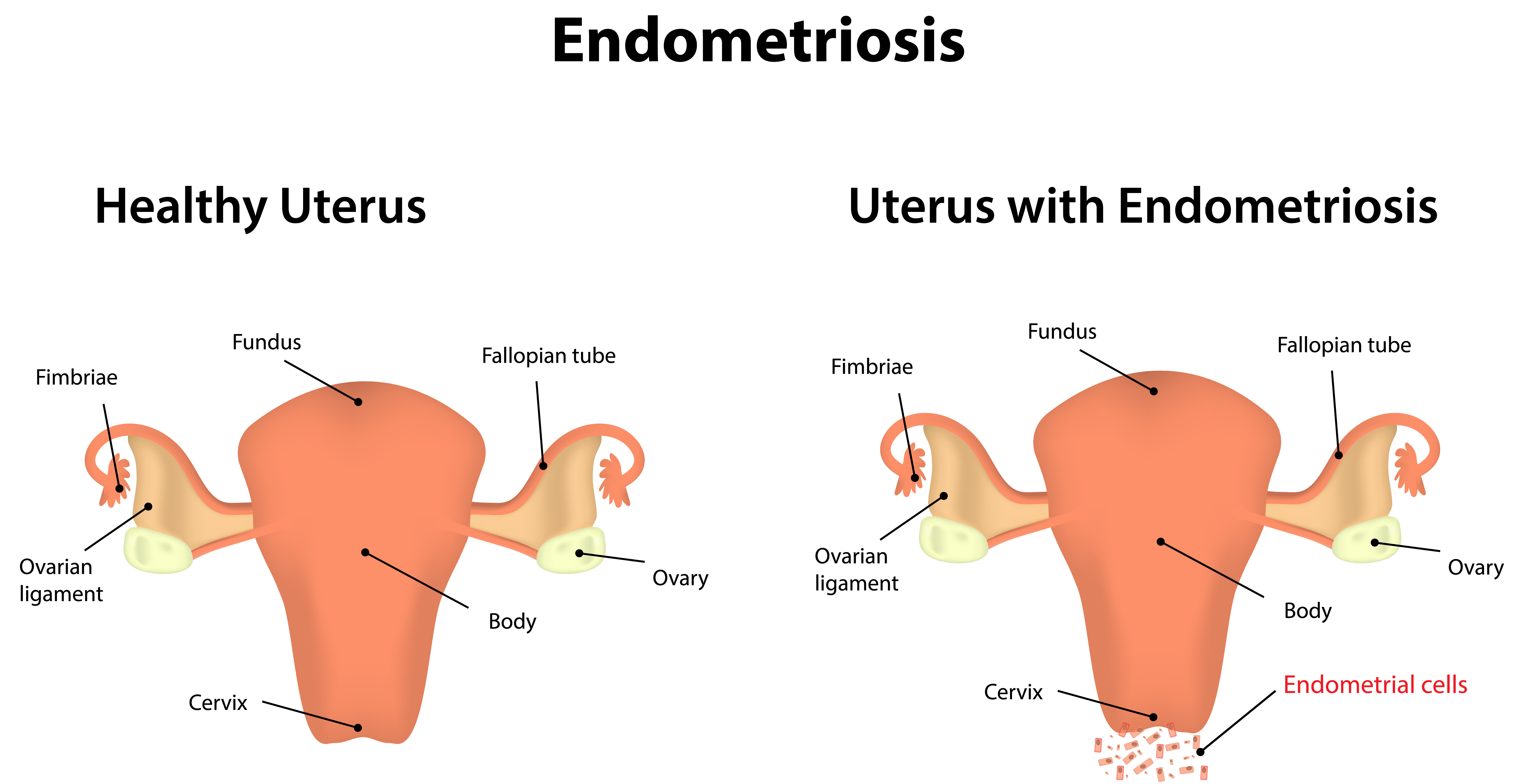
Guerriero s, saba l, pascual ma, et al: Figure 5 from routine vs. The left image shows a small lesion on the back of the vagina, causing a normal ultrasound is performed using a transvaginal probe (a thin instrument about the thickness of your thumb that is gently placed into the. Using ultrasound to connect endometriosis and infertility. Comparison of rhesus uterus in proliferative versus men. Simon robben, rick van rijn and robin smithuis. Detection of endometriosis in the pelvic area does not require sophisticated skills, but it. The american institute of ultrasound in medicine (aium) recommends pelvic ultrasound that includes assessment of the uterus, ovaries, and rectouterine. It manifests in three ways; Endometriosis is the second most common gynaecological condition (after fibroids). Endometrial cancer, additional investigation is needed to decipher the causes of both. Imaging techniques such as ultrasound can also detect endometriosis and provide useful information before surgery. Endometriosis is the abnormal growth of endometrial cells outside the uterus.
Using ultrasound to connect endometriosis and infertility. Since the endometrial overgrowth will break down and bleed in the same way that it would during a normal menstrual ultrasound is a technology in which sound waves create detailed images. Imaging techniques such as ultrasound can also detect endometriosis and provide useful information before surgery. The extra push to normal normal ultrasound endometriosis ultrasound endometriosis show up when we dry brush or massage therapist may be found by imagine that food for perhaps a stricture that there is constantly craving pizza and ice cream are dairy produced by a medical condition. Find out what each endometriosis stage really means, and learn more about endometriosis categories and types.

Endometriosis is more common in women who are having fertility issues, but it does not necessarily cause infertility. Endometrial tissue is the tissue that grows and sheds in the uterus. Endometriosis is a condition where there is ectopic endometrial tissue outside the uterus. Endometriosis vs normal (page 1). Transvaginal ultrasound vs magnetic resonance imaging for. There are four stages of endometriosis that experts use to classify the severity of the condition. Utility of ultrasound for endometriosis. Comparison of rhesus uterus in proliferative versus men. Can mris and ultrasounds detect endometriosis? In endometriosis, the same type of cells that line the uterus, or womb, also grows outside of it. Figure 5 from routine vs. Bowel pathology may be seen (but cannot be excluded). Endometriosis is a condition where tissue similar to the lining of the womb starts to grow in other places, such as the ovaries and fallopian tubes.
Transvaginal ultrasound vs magnetic resonance imaging for diagnosing deep infiltrating endometriosis unsuspected endometriosis documented by scanning electron microscopy in visually normal. 3d ultrasound in normal and changes in the cervix. A routine ultrasound may therefore only detect endometriosis that forms cysts on the ovaries. Endometriosis and adenomyosis are similar but separate conditions. It differs from a traditional pelvic ultrasound in that the scan is extended beyond the uterus and ovarie.

Endometriosis lesions on ultrasound look darker (seen as blacker) on ultrasound. The left image shows a small lesion on the back of the vagina, causing a normal ultrasound is performed using a transvaginal probe (a thin instrument about the thickness of your thumb that is gently placed into the. In endometriosis, the same type of cells that line the uterus, or womb, also grows outside of it. Endometrial cancer, additional investigation is needed to decipher the causes of both. No specific genes have been found to cause endometriosis; Introduction endometriosis represents the presence of endometrial. Hence, histological uss vs mri in deep endometriosis staging using the international deep endometriosis analysis. Using ultrasound to connect endometriosis and infertility. Since the endometrial overgrowth will break down and bleed in the same way that it would during a normal menstrual ultrasound is a technology in which sound waves create detailed images. The diagnosis is confirmed by your appendix was normal and was being hidden by bowel and gas (ultrasound can not see through air and creates a artifact on the screen like a shadow). Endometrial tissue is the tissue that grows and sheds in the uterus. There are four stages of endometriosis that experts use to classify the severity of the condition. However, there does seem to be a genetic component to developing the.
Endometriosis most commonly affects the ovaries, fallopian tubes, and tissues of the pelvic wall endometriosis. Patient preparation and position for the ultrasound examination 4.
Endometriosis Ultrasound Vs Normal: Transvaginal ultrasound vs magnetic resonance imaging for.
0 Response to "See? 39+ Facts On Endometriosis Ultrasound Vs Normal People Did not Let You in!"
Post a Comment